

Abstract
Chemical modification of silicon nitride (SiN) microsieves with glutaraldehyde and 3-glycidoxypropyldimethylethoxysilane (GOPS) for bio-coupling with an antibody to capture MCF-7 circulating tumor cells of breast cancer is reported. In this research, the antibody monoclonal anti-cytokerantin–FITC with fluorescein isothiocyanate label was used due to its good selectivity to MCF-7 circulating tumor cells of breast cancer. Modification efficiency was determined by the variation of contact angle. The increase in contact angle of the microsieves treated with glutaraldehyde and GOPS indicated that the microsieve surface changed from hydrophilic to hydrophobic. These results confirmed the successful immobilization of glutaraldehyde and GOPS onto SiN microsieves. Antibody binding effect was evaluated by fluorescence microscopy. Fluorescent images exhibited that GOPS was more effective than the glutaraldehyde treatment. The GOPS-treated microsieves were then used for capture of MCF-7 cells in phosphate buffered saline (PBS). The fluorescent images proved that the surface modification of SiN microsieves with GOPS helped to increase the efficiency of MCF-7 capture.
Export citation and abstract BibTeX RIS

Original content from this work may be used under the terms of the Creative Commons Attribution 3.0 licence. Any further distribution of this work must maintain attribution to the author(s) and the title of the work, journal citation and DOI.
1. Introduction
Breast cancer, one of the most popular diseases in recent years, killed over 40 000 people in the United States in 2014 [1] and about 1.7 million new cases are found each year all over the word. Breast cancer is sometimes found after symptoms appear, but many women with early breast cancer have no symptoms. In Vietnam, 70% of patients have been at a late stage before medical examination, whereas 80% of patients have been saved thanks to early detection and timely treatment [2]. If breast cancer could be detected early, many human lives could be saved. This shows us the importance of screening tests before any symptoms develop.
Circulating tumor cells (CTCs) were first known by an Australian doctor Thomas Ashworth in 1869. The CTCs are the cells which are generated and separated from the primary or secondary tumors and intrude into the circulatory system through thin capillaries. The presence of CTCs is not synonymous with the formation of metastases. Assessment of CTCs may be useful in monitoring and treatment process and their characteristics can help to develop new treatments to prevent progression of metastatic cancer [3]. However, detection of CTCs is very difficult because the fraction of CTCs on whole blood cells is very low (about 1:107 to 1:108) [4]. Therefore, many methods have been developed to capture and detect the CTCs such as di-electrophoresis, molecular sieves, centrifugation, optical spectrum and immunoaffinity methods [5].
Microsieve membranes fabricated by micro-technology have been used in microfiltration technology as a new generation of inorganic microfiltration membranes. The microsieve membranes fabricated from silicon nitride have excelent properties including being chemically inert and having high mechanical strength. Ultra-thin film thickness may reach 150 nm with uniform pore size, shape and high porosity. Thanks to good performance and high selectivity, microsieves membranes have been widely used in biological filtration, diagnosis and bacteria testing. In principle, the membranes, which have a pore size smaller than the size of bacteria, can be used to filter and detect bacteria in a biological fluid and to eliminate other components easily. However, a fouling problem due to accumulation of biological particles in the filtering process has seriously affected applications of microsieves. One of the most feasible solutions is chemical treatment of the membrane surface to reduce the accumulation and enhance biological selectivity of the membrane. Many studies have been done using surface chemistry approaches for bio-receptor attachment on silicon-based materials [6–12].
Table 1 shows the agents which can crosslink with silicon nitride (SiN) and silicon oxide (SiOx) surfaces [13]. The coupling agents must be di-function compounds, the former is used for binding substrate and the latter is used for binding bio-receptor. For instance, the SiOx surface aminated by amino-organsilane was used to immobilize antibody by absorption or carboxylate before antibody immobilization. Thiol-ended organosilane was employed to mount on surface and immobilize antibody via disulfide bonds. Epoxy-organosilane has reactivity to nucleophiles groups such as amine, thiol, hydroxy. The conjunction between carboxyl-organosilane and proteins must be added with 1-ethyl-3-(3-dimethylaminopropyl) carbodiimide/N-hydroxysulfosuccinimide (EDC/NHS). Isocyanate-ended organosilane compounds react with amine media to form urea bonds and with hydroxyl media to form urethane bonds. Reaction of SiH with alkene must be done in thermal or photochemical activation. Organosilane compounds are used for SiOx in preference to SiN. Using organosilane compounds for modified SiN is rare unless SiN has been oxidized previously.
Table 1. Some crosslink agents used for bio-receptor attachment on silicon-base materials.
| Substrate | Coupling agents | Substrate-link | Bio-link | Comments |
|---|---|---|---|---|
| -Si-OH | Amino-organosilane | Siloxan | Amino absorption, other agents (di-carboxylic) | Weak complicate |
| -Si-OH | Thiol-organosilane | Siloxan | Thiol bridge, other agents (alken, acid...) | Complicate |
| -Si-OH | Epoxy-organosilane | Siloxan | Amino, imide, thiol | Multiple choice |
| -Si-OH | Carboxyl-organosilane | Siloxan | Amide, ester | Simple |
| -Si-OH | Isocyanate-organosilane | Siloxan | Urethane | Hard control |
| -Si-NH2 | Di-aldehyde | Imide | Imide | Simple |
| -Si-H | Alkene-carboxylic | Silane | Amide | UV lamp |
In this study glutaraldehyde and 3-glycidoxypropyldimethylethoxysilane (GOPS) were employed for chemical modification of SiN surface because of simple procedure and strong adhesion with both substrate and bio-receptors.
MCF-7 is a common commercial cell which has been used in breast cancer research with the size of 20–24 μm. In our research, MCF-7 was used to represent CTCs because of its popularity and the antibody monoclonal anti-cytokerantin–FITC with high selectivity affinity to MCF-7 was applied.
In this paper we report the chemical modification of SiN substrate with glutaraldehyde and 3-glycidoxypropyldimethylethoxysilane (GOPS) for bio-coupling with the antibody monoclonal anti-cytokerantin–FITC to capture MCF-7. The antibody monoclonal anti-cytokerantin–FITC with the green light of the fluorescence indicator FITC will indicate if the modification reaction is successful (figure 1).

Figure 1. Illustration of surface modification of SiN microsieves.
Download figure:
Standard image High-resolution image2. Experimental
2.1. Materials
The MCF-7 breast cancer cells, monoclonal anti-cytokerantin–FITC antibody, glutaraldehyde 50%, 3-glycidoxypropyldimethylethoxysilane (GOPS) 97%, sodium periodate and sodium cyanoborohydride were purchased from Sigma-Aldrich. HF (40%), H2SO4 (96%), H2O2 (30%) and acetone were purchased from Merck, Germany. Sodium phoshatedibasic, sodium phosphate monobasic, citric acid, sodium citrate and acetic acid were bought from China.
Phosphate-buffered saline (PBS) pH 7.5 was prepared from sodium phosphate monobasic 0.032 M and sodium phosphate dibasic 0.168 M. Citrate buffer pH 5.4 was prepared from citric acid 0.016 M and sodium citrate 0.034 M.
2.2. Experimental
2.2.1. Modification by glutaraldehyde
Firstly, the SiN wafer was washed with acetone, dichloromethane and dried in air. Then the wafer was treated with a piranha solution (H2SO4:H2O2 = 2:1) for 5 min to eliminate impurities on the surface. Subsequently, the activation process of the SiN substrate was performed in a two-neck round bottom flask with HF 1% solution for 3 min under nitrogen atmosphere and then the wafer was washed 3 times with PBS (pH 7.5). Then, the wafer was soaked in PBS solution containing 5% glutaraldehyde under nitrogen for 2 hours at room temperature. After that the wafer was cleaned with PBS and DI water for 5 min and dried in air (figure 2). After glutaraldehyde modification process, the wafer was incubated with monoclonal anti-cytokerantin-FITC solution (50 μg ml−1) for 2 h in a dark chamber at room temperature. Finally, the wafer was washed with PBS and de-ionized water and then dried in air. Fluorescent images were recorded by fluorescence correlation microscopy BX41 (Olympus, Japan).

Figure 2. Schemes of SiN surface modification by glutaraldehyde.
Download figure:
Standard image High-resolution image2.2.2. Modification by GOPS
The SiN substrate was treated with the piranha solution for 5 min to activate the surface before modification. The silanized surface was treated with a solution of 3% 3-glycidoxypropyldimethylethoxysilane (GOPS) acidified to pH 2 with sulfuric acid 1 M overnight at 50 °C. The oxidation of GOPS group to aldehyde groups was performed by using sodium periodate 0.5% in glacial acetic acid 80% at room temperature (figure 3). Monoclonal anti-cytokerantin-FITC was fixed on GOPS substrate and GOPS oxidized substrate through the deamination reaction for 15 min at room temperature by using 0.5 mg ml−1 protein and 10 mg ml−1 in 100 mM sodium cyanoborohydride buffer citrate pH 5.5.

Figure 3. Schemes of SiN surface modification by GOPS.
Download figure:
Standard image High-resolution image2.2.3. Characterization of SiN substrates after the treatment steps
The SiN surface is pretty chemically inert and hydrophobic, and the contact angle of virginal SiN surface is usually 36° [13]. After modification with aldehyde and epoxy groups, the SiN surface will change the wetting properties from hydrophobicity to hydrophilicity and the water contact angle will be smaller. This is the easiest method to identify whether the process is successful or not. The hydrophobic or hydrophilic property of the SiN surface was determined by contact angle. The contact angle instrument CAM 101 (KSV Instruments, Finland) was used after each step of surface treatment.
Thickness of the organic layers was determined by Smart SE Spectroscopic Ellipsometer (Horiba Scientific, France). Finally, the fluorescence microscope BX41 (Olympus, Japan) was used to detect if the antibody was attached to the SiN surface via the coupling agents such as aldehyde, epoxy. This is the most observable method to evaluate the coupling efficiency and performance on the SiN substrates.
3. Results and discussion
3.1. Contact angle measurement
After treatment with glutaraldehyde and GOPS, the wetting properties of SiN surface changed dramatically. The results of the contact angle measurement of SiN surface are presented in figure 4 and table 2.

Figure 4. Contact angle values of the SiN surface after the modification and treatment steps with glutaraldehyde, GOPS and oxidized GOPS.
Download figure:
Standard image High-resolution imageTable 2. Contact angle values of the SiN surface after the modification steps.
| Coupling agents | After cleaning surface | After surface modification | After antibody immobilization |
|---|---|---|---|
| Glutaraldehyde 5% | 36.83° ± 5.89° | 20.37° ± 6.63° | 34.97° ± 3.66° |
| GOPS 3% | 22.28° ± 2.50° | 35.62° ± 2.14° | 46.95° ± 1.35° |
| GOPS 3% oxidized | 22.28° ± 2.50° | 78.98° ± 6.42° | 85.96° ± 3.56° |
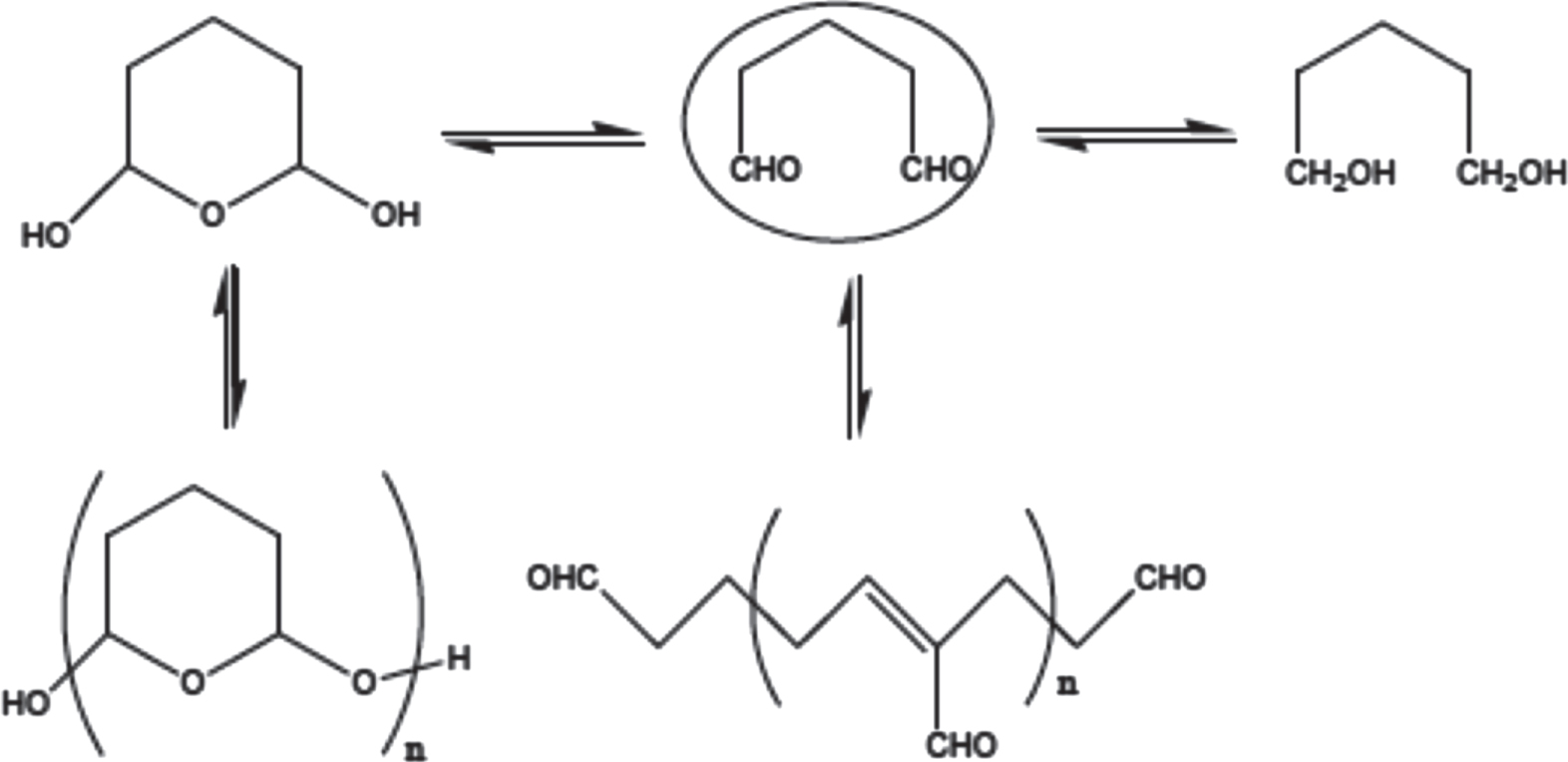
After the treatment with glutaraldehyde 5%, the contact angle of the surface significantly reduced to 20.37°. However, when the antibodies were attached to the surface, the contact angle increased again to 34.97° and approximately reached the value before the glutaraldehyde modification. This result contradicts the predictions and the results of Bañuls et al [7]. It was assumed that the reactivity of aldehyde functions at both sides was equal (figure 5) or glutaraldehyde might exist in other conformations (figure 6) [14–16], as the antibody might not be attached successfully. More methods are needed to confirm the results of glutaraldehyde treatment.

Figure 5. Formation of di-imide on the SiN substrate of glutaraldehyde.
Download figure:
Standard image High-resolution image
Figure 6. The other conformations of glutaraldehyde.
Download figure:
Standard image High-resolution imageOn the other hand, when the substrate was modified with GOPS and activated GOPS, the contact angle increased significantly. These results can be explained by the fact that GOPS has hydrophobic hydrocarbon chain which should be attached to the SiN surface. The chain makes the hydrophobicity of the surface increase and increase the contact angle of the SiN substrate. In addition, after each treatment step, the refractive coefficient of the SiN surface changed (figure 7).

Figure 7. The change of the refraction of the SiN surface after treatment with (a) GOPS; (b) with GOPS linked to antibody; (c) oxidized GOPS.
Download figure:
Standard image High-resolution image3.2. Thickness measurement
Besides the contact angle measurement, ellipsometry was used to determine the thickness of the layers by simulating and calculating the change of the parameters including reflection angle and refractive index of the surface. Figure 8 shows the model for thickness measurement by ellipsometry.

Figure 8. Model used in the ellipsometer charaterization.
Download figure:
Standard image High-resolution imageThickness measurement results of the SiN substrate modified by glutaraldehyde are shown in table 3. The results show that the SiN layer thickness before modification was about 900 nm, which matched with the thickness declared by the vendor (1000 nm). After glutaraldehyde attachment, the thickness of glutaraldehyde layer was about 50 nm, thicker than a single layer of glutaraldehyde reported by Bañuls et al [7] (about 3 nm). Therefore, the glutaraldehyde layer is supposed to be in oligomer state.
Table 3. Layers thickness calculation results of the glutaraldehyde treatment.
| Before modification | After treatment with glutaraldehyde 5% | After antibody binding | |
|---|---|---|---|
| SiN layer thickness (L1) | 895.5 nm | 821.1 nm | 505.2 nm |
| Organo layer thickness (L2 + L3) | 0 nm | 51.9 nm | 375.9 nm |
After the attachment of the antibodies, the thickness of the organic layer increased to 380 nm, but the thickness of the SiN layer decreased to 500 nm. Similar to the case of GOPS and oxidized GOPS, the GOPS thickness was 0.19 nm (table 4) which is quite small in comparison with the size of single molecules of GOPS. After the antibodies attachment, the thickness of the organic layer was more than 800 nm, which was quite different from the GOPS layer. This error may be caused by the lack of database of the organic layer antibody used in the experiments. Moreover, the rough of the GOPS layer caused scattering of the light irradiation on the inaccurate obtained light signal. The thickness of the oxidized GOPS layer was about 20 nm and the SiN layer was about 900 nm, which is more accurate due to a less rough surface.
Table 4. Layers thickness calculation results of the treatment with GOPS and oxidized GOPS.
| After cleaning | After GOPS treatment | After antibody binding on GOPS substrate | After oxidized GOPS treatment | |
|---|---|---|---|---|
| SiN layer thickness (L1) | 895.5 nm | 721.3 nm | 813.60 nm | 912.19 nm |
| Organo layer thickness (L2 + L3) | 0 nm | 0.19 nm | 856.10 nm | 20.72 nm |
The contact angle and ellipsometry results indicated that the SiN substrate modification with glutaraldehyde and GOPS was successful. However, the mechanism and layer thickness of these treatments must be studied further.
3.3. Fluorescence microscopy
Figure 9 shows the fluorescent microscopy images of the SiN substrates treated with glutaraldehyde before and after immobilization of the antibody fluorescent markers monoclonal anti-cytokerantin-FITC. After treatment with glutaraldehyde, the substrates were free with fluorescent. But after the antibody mounting, tiny bright spots appeared on the substrate, as shown in figure 9(b). In comparison with the GOPS treatment, the fluorescent image showed large green spots spreading on the substrate surface after antibody soaking as shown in figure 10(b).

Figure 9. Fluorescent microscopy images of the SiN substrate treated with glutaraldehyde. (a) Before mounting antibody, (b) after mounting antibody.
Download figure:
Standard image High-resolution image
Figure 10. Fluorescent microscopy images of the SiN substrate treated with GOPS. (a) Before mounting antibody, (b) after mounting antibody.
Download figure:
Standard image High-resolution imageSimilar results were obtained for the substrate treated with the activated GOPS after soaking in antibody (figure 11). In this image, the fluorescent zones glowed continuously. It should be due to the effect of the activating GOPS before the antibody was attached, which made the fluorescence become homogenous.

Figure 11. Fluorescent microscopy image of the SiN substrate treated with oxidized GOPS and soaked in the antibody solution.
Download figure:
Standard image High-resolution image
4. Conclusion
Chemical modification of silicon nitride substrate with glutaraldehyde and GOPS was successful. The fluorescence microscopy images showed that the mounting antibody on glutaraldehyde treatment surface was not effective but the treatment with GOPS and oxidized GOPS was much better. The results showed the mounting antibody on the oxidized GOPS treated surface became more homogenous. This surface treatment could be applied for the microsieves to capture MCF-7 circulating tumor cells of breast cancer.
Acknowledgments
The authors would like to thank Ministry of Science and Technology of Vietnam for funding this research.