

Abstract
A simple chemical reduction method is used to prepare colloidal bimetallic Cu–Ag core–shell (Cu@Ag) nanoparticles. Polyvinyl pyrrolidone (PVP) was used as capping agent, and ascorbic acid (C6H8O6) and sodium borohydride (NaBH4) were used as reducing agents. The obtained Cu@Ag nanoparticles were characterized by powder x-ray diffraction (XRD), transmission electron microscopy (TEM) and UV–vis spectrophotometry. The influence of [Ag]/[Cu] molar ratios on the formation of Ag coatings on the Cu particles was investigated. From the TEM results we found that the ratio [Ag+]/[Cu2+] = 0.2 is the best for the stability of Cu@Ag nanoparticles with an average size of 22 nm. It is also found out that adding ammonium hydroxide (NH4OH) makes the obtained Cu@Ag nanoparticles more stable over time when pure deionized water is used as solvent.
Export citation and abstract BibTeX RIS

Original content from this work may be used under the terms of the Creative Commons Attribution 3.0 licence. Any further distribution of this work must maintain attribution to the author(s) and the title of the work, journal citation and DOI.
1. Introduction
One of the important trends in microelectronic back-end processes is the application of metallic nanoparticle (NP) suspensions or pastes, which have been widely used as conductive inks (namely metallic inks) to manufacture fine-pitch electrical line patterns for organic transistors, radio frequency identification (RFID) antennas, or ultra large scale integration (ULSI) interconnects not only because of their high electrical conductivity and flexibility in handling, but also the low processing temperature [1, 2]. The reduced processing temperature is due to the large surface-to-volume ratio of the particles leading to a dramatic lowering of the melting point and sintering transition. Ag nanoparticles are most commonly used for metallic inks due to the mature synthesis techniques and excellent performance. In order to cut the material cost, Cu nanoparticles have been considered for some time as a replacement for Ag nanoparticles in nanoparticle-based interconnect applications [3, 4]. Cu has the advantages of excellent electrical conductivity (only 6% less than that of Ag) and much lower price. However, nanocopper oxidizes rapidly under ambient conditions. To fabricate a conductive Cu pattern on a plastic substrate (polyimide), Cu nanoparticles usually have to be heated at 200 °C under a reductive atmosphere to remove surfactants and thereby obtain acceptable electrical conductivity. When used as bonding materials, Yan et al demonstrated that the joints created with Cu nanoparticles yield a low resistivity (86 μΩ cm) after sintering at 300 °C in air under a bonding pressure of 5 MPa [5]. Such joints can be applied as die-attach materials for high power chips for automotive electronics or high power devices, frequently working at a temperature of the order of 200 °C or above. Therefore, it is crucial to improve the stability of Cu nanoparticles to render these inks practical.
Several strategies have been proposed to improve the stability of Cu nanoparticles against oxidation. Non-oxidizable coatings of carbon, ligands, polymers, silica and noble metals have been suggested [4, 6, 7]. Cu nanoparticles with Ag coatings appear promising for interconnect applications because they possess high electrical conductivity. The Ag shells are able to act as a connector between Cu particles and assist sintering [7].
There are many ways to form Cu–Ag core–shell (Cu@Ag) nanoparticles such as electroplating, electroless plating, vacuum process, sputtering etc [5, 7, 8]. In this paper, a simple chemical reduction method was used to synthesize Cu@Ag nanoparticles using polyvinyl pyrrolidone (PVP) as an efficient protective agent in a one-step process. Ascorbic acid and sodium borohydride (NaBH4) are chosen as the reducing agents, due to their nontoxicity and easy availability. Cu@Ag nanoparticles powders prepared by this method easily disperse and hardly oxidize even after a long time.
2. Experimental
2.1. Materials
All chemicals were used without further purification. Copper (II) sulfate pentahydrate salt (CuSO4.5H2O, Merck) has 98.0% purity. Silver nitrate (AgNO3, Merck) and polyvinyl pyrrolidone (PVP, average molecular weight of 40 000, BASF) were used as capping agents. Sodium borohydride (NaBH4—Reagent Plus 99%, Sigma-Aldrich) and ascorbic acid (99.7%, Prolabo) were used as the reducing agents. Sodium hydroxide NaOH (>98%, China) was used to adjust the pH and ammonium hydroxide (NH4OH, Merck) was also used to dissolve silver nitrate and copper sulfate pentahydrate.
2.2. Synthesis of Cu@Ag nanoparticles
Polyvinyl pyrrolidone (PVP) 40 000 and ascorbic acid are first separately dissolved in deionized water. Then the two solutions are mixed and stirred at 50 °C. Copper (II) sulfate pentahydrate salt, CuSO4.5H2O (0.01 M), and silver nitrate, AgNO3, are separately dissolved in NH4OH to obtain complex ions [Cu(NH3)4]2+ and [Ag(NH3)2]+. A solution of NaBH4 is poured into the stirring solution. [Cu(NH3)4]2+ and [Ag(NH3)2]+ solutions are then dropped one after the other into the first solution. The temperature is kept at 50 °C during the synthesis.
3. Results and discussion
In this paper the action of ascorbic acid (C6H8O6) and sodium borohydride (NaBH4) used as reductants in reducting the metal salts CuSO4 and AgNO3 is shown through the following reactions [9–12]


Besides, the chemical reduction reaction of Ag ions involves Cu atoms already present in solution.
3.1. Influence of the ratio [Ag+]/[Cu2+]
Because Cu atoms are also consumed in the reduction of Ag ions as above [13], the ratio [Ag+]/[Cu2+] is an important factor influencing the formation and improvement of Cu@Ag nanoparticles. Therefore, we synthesized the samples with different [Ag+]/[Cu2+] ratios with all other ratios fixed as shown in table 1.
Table 1. Parameters for the Cu@Ag nanoparticles preparation reactions.
| Sample | [Ag+]/[Cu2+] | [Cu2+]/[C6H8O6] | [Cu2+]/[PVP] | [NaBH4]/[Cu2+] |
|---|---|---|---|---|
| B1 | 0.2 | 0.2 | 0.02 | 0.2 |
| B2 | 0.4 | 0.2 | 0.02 | 0.2 |
| B3 | 0.6 | 0.2 | 0.02 | 0.2 |
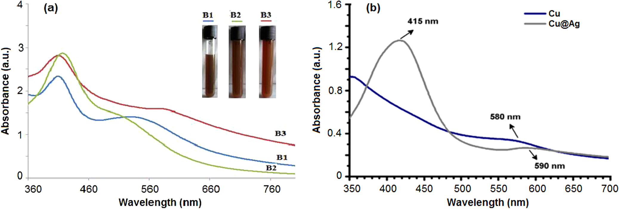
Figure 1(a) shows that the colors of the three samples are nearly identical; the solution color tends to be darker when the volume of Ag+ increases. The samples have two absorption peaks. The first peak appears at a wavelength of about 410 nm known as the typical aborption peak position of Ag nanoparticles [10, 14]. The second peak appears at a wavelength from 525 nm to 580 nm, known as the absorption peak position of Cu nanoparticles [15]. As shown in figure 1, the spectra for the three samples are similar to the absorption spectrum of Cu@Ag nanoparticles published by a group of researchers from Taiwan National University of Science and Technology [14]. For sample B3, the second absorption peak is shifted to longer wavelengths, closer to the absorption peak position of Cu oxide. We assume that this sample has oxidized Cu nanoparticles.

Figure 1. (a) UV–vis spectra and photographs of B1, B2, B3 samples, and (b) UV–vis spectra of Cu@Ag nanoparticles of a research group from Taiwan National University of Science and Technology.
Download figure:
Standard image High-resolution imageFigure 2 shows that there are core–shell bimetallic nanoparticles and non core–shell metallic nanoparticles in sample B1 and B2. We assume that the non core–shell nanoparticles are Ag and Cu nanoparticles in solution. The size of Cu@Ag nanoparticles in sample B1 is smaller than in sample B2 and from 15 nm to 22 nm. The size of core–shell Cu@Ag particles in B2 is larger and about 35 nm. Sample B2 with a larger volume of Ag+ contains more Ag atoms making nanoparticles in sample B2 reach a larger size than in sample B1. Moreover, in the reduction reaction of Ag+ ion using Cu0 atoms, Cu2+ ions are put back in solution after reaction. These Cu2+ ions will continue to be reduced by ascorbic acid and sodium borohydride and become Cu0 atoms which are involved in the improvement.

Figure 2. (a) TEM images and (b) particle size distributions of samples B1 and B2, respectively.
Download figure:
Standard image High-resolution image3.2. Influence of NH4OH solution and deionized water
As described above, we use NH4OH solution to dissolve CuSO4 and AgNO3 in order to create complex ions [Cu(NH3)4]2+ and [Ag(NH3)2]+. We also replaced NH4OH solution with deionized water in order to dissolve CuSO4 and AgNO3 to lead to Ag+ and Cu2+ ions.
Samples C1 and C2 are synthesized with the same reaction preparation parameters as samples B1 and B2 but the NH4OH solution is replaced by deionized water.
Figure 3 shows the appearance of Cu@Ag nanoparticles in both samples, with an average particle size from 30 to 40 nm. The sizes of Cu@Ag nanoparticles of sample C1 and C2 are larger than sample B1 and B2. After monitoring the samples over time, agglomerates started to appear at the bottom of the vials after 12 days, but no agglomerates appeared in sample B1 and B2. We suppose that the reduction of complex ions [Cu(NH3)4]2+ and [Ag(NH3)2]+ creates particles with better stability than with Ag+ and Cu2+ ions.

Figure 3. (a) TEM images and (b) particle size distributions of samples C1 and C2, respectively.
Download figure:
Standard image High-resolution imageFigure 4 shows that sample B1 still has two absorption peaks after 80 days from preparation in the ranges 1 and 2. Under visual observation there is no agglomerate at the bottom of the vial after 80 days of preparation. Sample B2 presents a small quantity of agglomerate after 80 days. The sample B1 ([Ag+]/[Cu2+] = 0.2) presents the best stability over time. We continue to observe its stability.

Figure 4. UV–vis spectra of sample B1 at different times.
Download figure:
Standard image High-resolution imageFigure 5 shows that sample B1 has diffraction peaks at the positions of Cu and Ag. Interestingly we do not observe diffraction peaks of Cu oxide, contrary to what is reported in reference [14]. This leads us to think that the volume of Cu oxide is either null or very small.

Figure 5. X-ray diffraction of sample B1.
Download figure:
Standard image High-resolution image
4. Conclusion
In this paper Cu@Ag nanoparticles are successfully synthesized using a chemical reduction method. The particles present an average size of about 22 nm and their stability is longer than 80 days. A ratio of [Ag+]/[Cu2+] = 0.2 is the best among the three tested for the stability of Cu@Ag nanoparticles. Using ammonium hydroxide (NH4OH) as solvent also improves the stability of the obtained Cu@Ag nanoparticles over time as compared with deionized water. According to the XRD measurement, there is no appearance of Cu oxide in the samples.
Acknowledgments
The authors greatly appreciate the financial support of the Ministry of Sciences and Technology of Vietnam.